Series: Well Woman Conversations
By: Dr Raman Narang, Consultant Medical & Hemato-Oncologist
Last reviewed: September 2026

The direct answer
If you are 40 or older, have no breast symptoms and are at average risk, mammography is the main breast screening after 40 test to discuss with your doctor. Major guidelines agree that mammography has an important role from the 40s onward, although they differ on whether it should be done every year or every two years. Ultrasound is not usually a replacement for mammography, while breast MRI is generally reserved for women whose risk is substantially higher than average.
Turning 40 often comes with a long list of health checks. Breast screening can be particularly confusing because women hear very different advice: “Do a mammogram every year,” “You only need an ultrasound,” “Wait until 50,” or “MRI is more accurate, so why not just do that?”
The correct answer is not simply to order every available scan.
The first question is what is your breast cancer risk? The second is whether you are being screened while well or investigated because you have a symptom. Those two situations require different approaches.
I’m healthy and 40. Do I need a mammogram now?
For many women at average risk, age 40 is now an appropriate point to begin a conversation about regular mammography.
The US Preventive Services Task Force recommends mammography every two years from ages 40 to 74. ACOG recommends beginning mammography at 40, with screening every one or two years after discussion with the patient. The American College of Radiology recommends annual mammography starting at 40. The American Cancer Society gives women aged 40–44 the option of annual screening and recommends annual mammography from 45–54.
There is also genuine professional debate. Updated American College of Physicians guidance published in 2026 recommends shared decision-making for average-risk women aged 40–49; women who choose screening may undergo mammography every two years. It recommends biennial mammography from 50–74.
So if two well-informed doctors recommend slightly different screening intervals, it does not automatically mean one is wrong. Different guidelines weigh the additional cancers detected by more frequent screening against false-positive results, additional tests, biopsies, anxiety and overdiagnosis differently.
What does “average risk” mean?
Broadly, average risk means there is no known major factor that places you in a substantially higher-risk category—for example:
- a known inherited breast-cancer predisposition such as a pathogenic BRCA1 or BRCA2 variant;
- a strong pattern of breast or ovarian cancer in the family;
- previous therapeutic radiation to the chest at a young age;
- a previous breast cancer; or
- certain high-risk abnormalities found on a previous breast biopsy.
A woman can feel completely healthy and still warrant a more detailed risk assessment.
Should I have a mammogram every year or every two years?
For an average-risk woman in her 40s, both annual and biennial strategies appear in respected guidelines.
Annual screening means more opportunities to detect a cancer between scheduled examinations, but it also exposes a woman to more screening episodes and therefore more chances of a false-positive result, recall imaging or biopsy.
Biennial screening reduces some of these screening-related harms while still providing a demonstrated mortality benefit. The USPSTF currently recommends every two years from 40–74, while ACOG considers either a one- or two-year interval reasonable after shared decision-making.
For an individual woman, I would therefore avoid treating the interval as an isolated number. Her age, previous mammograms, breast density, family history and overall risk matter.
The most useful screening programme is also one that actually continues. A carefully chosen, documented schedule followed consistently is more valuable than repeatedly changing tests without a clear reason.
Is breast ultrasound enough after 40?
Usually, no.
Ultrasound and mammography provide different information. Mammography can identify changes—including certain calcifications—that may not be apparent on ultrasound. Ultrasound is particularly useful when evaluating a specific lump or mammographic abnormality and has other important diagnostic roles.
It may also be considered as supplemental imaging in selected women, including some women with dense breasts. However, current evidence has not established that routinely adding ultrasound after a negative mammogram improves long-term health outcomes for every woman with dense breasts. The USPSTF considers evidence insufficient to recommend routine supplemental ultrasound or MRI solely because breasts are dense.
The 2026 ACP guidance goes further for average-risk women with dense breasts, advising against routine supplemental screening MRI or ultrasound and suggesting that digital breast tomosynthesis can instead be considered after discussion of benefits, harms, availability and cost.
So an annual “breast ultrasound package” should not automatically be treated as equivalent to evidence-based mammographic screening.
My report says I have dense breasts. What does that change?
Dense breasts are common and are not a disease.
Breast density is determined from a mammogram. Dense glandular and fibrous tissue appears white on mammography, and many breast cancers also appear white. This can make a cancer more difficult to see. Breast density is also independently associated with increased breast cancer risk.
But “dense breasts” does not automatically mean “you need an MRI.”
Instead, density should trigger a broader question:
Dense breasts plus what other risk factors?
A woman with dense breasts but otherwise average risk is different from a woman with dense breasts, multiple close relatives affected at young ages and a previous high-risk breast biopsy.
That distinction is why breast density should be interpreted as part of a risk profile rather than in isolation.
Is 3D mammography better than a standard mammogram?
Digital breast tomosynthesis often called 3D mammography takes multiple low-dose images from different angles to construct a more detailed view of the breast.
Both conventional digital mammography and digital breast tomosynthesis are considered effective screening approaches by the USPSTF. Evidence reviewed by the NCI suggests tomosynthesis can improve detection and modestly reduce false-positive recalls compared with conventional digital mammography, particularly in some women with dense breasts.
That does not mean everyone must travel farther or pay considerably more simply to access a 3D machine.
If high-quality 3D mammography is readily available, it can be a reasonable option. If not, standard digital mammography remains an established screening method.
The quality of imaging, interpretation and appropriate follow-up matters at least as much as the label on the machine.
Who may need MRI or screening before the usual age?
This is where risk assessment becomes more important than age alone.
Women at substantially increased risk may require a different screening strategy, sometimes including both mammography and breast MRI and sometimes beginning before age 40.
Examples include women with certain inherited cancer-predisposition variants, a calculated lifetime breast cancer risk of around 20% or more, or a history of significant chest radiation at a young age. The ACR recommends earlier MRI surveillance for several such higher-risk groups, while the American Cancer Society similarly recommends combined MRI and mammography for selected women at high risk.
A family history should therefore not be reduced to the question, “Did your mother have breast cancer?”
The paternal side matters too, as do the ages at diagnosis and histories of ovarian, pancreatic, prostate and other relevant cancers within a family.
This is one reason cancer genetics and family-risk assessment can change a screening plan.
What if I already have a lump, nipple discharge or another breast change?
Then you are no longer discussing routine screening.
You need diagnostic evaluation.
Changes that should be assessed include a new breast or underarm lump, new nipple retraction, persistent or bloody nipple discharge, unexplained skin dimpling or thickening, or other persistent new breast changes. Indian ICMR clinical pathways similarly emphasise evaluation of breast lumps, nipple changes and suspicious skin changes.
The appropriate work-up may include clinical examination, diagnostic mammography, ultrasound and, where indicated, tissue sampling.
Do not wait for your “annual screening date” if a new symptom has appeared.
That distinction is important:
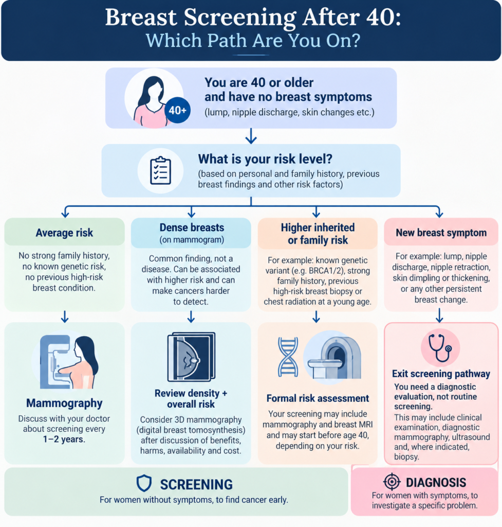
Screening is for women without symptoms. Diagnostic imaging investigates a problem that is already present.
What I look for clinically before recommending a screening plan
When discussing breast screening with a woman in her 40s, I am less interested in ordering the largest number of tests than in answering a few specific questions.
I look at:
- Her exact age and previous screening history — including whether earlier mammograms are available for comparison.
- Family history on both sides of the family — who developed which cancer and at what age.
- Personal breast history — previous biopsies, atypia, breast cancer or significant imaging abnormalities.
- Possible hereditary risk — whether the pattern warrants formal genetic counselling or testing.
- Previous chest radiation exposure, particularly at younger ages.
- Breast density, once mammography has established it.
- Whether she is genuinely asymptomatic — because a symptom changes the pathway from screening to diagnosis.
The objective is not simply to decide whether somebody needs “a scan.” It is to place her into the correct risk and diagnostic pathway.
Breast screening after 40: a simple if–then decision map
IF you are 40–74, have no symptoms and appear to be at average risk:
→ Discuss regular mammography. Depending on the guideline and your individual situation, this may be every one or two years.
IF you are 40–49 and unsure whether to start immediately:
→ Review your personal risk and the benefits and limitations of screening. Current professional guidelines differ, so shared decision-making is reasonable.
IF your mammogram reports dense breasts:
→ Do not simply replace mammography with ultrasound. Review the density finding together with your overall risk and discuss whether tomosynthesis or any supplemental imaging is appropriate.
IF you have a strong family history, inherited cancer-predisposition variant, previous high-risk lesion or relevant prior chest radiation:
→ Consider formal risk assessment. Your screening may need to begin earlier or include MRI alongside mammography.
IF you have a new breast symptom:
→ Do not treat it as a screening question. Arrange clinical and diagnostic evaluation.
IF you are over 75:
→ Screening decisions become increasingly individual and should consider overall health, life expectancy, previous screening and personal preferences.
A note for women in India
India’s National Health Mission public-health programme recommends breast screening by a trained provider for women from age 30, using periodic clinical breast examination, generally at least once every five years. This is designed for population-level screening within the Indian health system.
That programme should not be confused with every individual woman’s mammography decision.
WHO similarly distinguishes organised population-screening programmes from early diagnosis and notes that mammographic screening programmes depend on healthcare resources and the ability to ensure diagnostic follow-up and treatment.
For an individual woman with access to appropriate imaging, the practical decision should therefore combine her age, personal risk, family history and available high-quality mammography, rather than relying on a screening camp or imaging package alone.
Your breast-screening checklist after 40
Before your next preventive health visit, check these seven points:
- Know when your last mammogram was performed and keep the report and images if possible.
- Ask whether your current schedule should be annual or every two years.
- Read your mammography report for your breast-density category.
- Write down breast, ovarian and other relevant cancers on both your mother’s and father’s sides of the family, including the age at diagnosis.
- Mention any previous breast biopsy, atypical lesion, cancer or chest radiation.
- Do not substitute an ultrasound for mammography simply because ultrasound feels easier or avoids X-rays.
- Report any new lump, nipple or skin change promptly rather than waiting for routine screening.
Frequently asked questions
1. Can I do breast ultrasound instead of mammography after 40?
For an asymptomatic average-risk woman, ultrasound is generally not considered a replacement for screening mammography. It is commonly used to investigate specific findings and may occasionally supplement mammography in selected women.
2. Is the radiation from mammography dangerous?
Mammography uses a low dose of ionising radiation. Radiation exposure is one of the harms considered when guidelines evaluate screening, but major professional bodies conclude that the benefits of appropriately performed mammographic screening outweigh these harms for the age groups they recommend screening.
3. If I have no family history, do I still need screening?
Yes, absence of a family history does not place someone at zero risk. Age alone contributes to breast cancer risk, which is why screening recommendations also apply to average-risk women without affected relatives.
4. Do dense breasts automatically mean I need MRI?
No. Density can both increase breast cancer risk and make mammography more difficult to interpret, but supplemental MRI is not automatically recommended solely because breasts are dense. The decision depends on the woman’s overall risk profile.
5. Should I choose 2D or 3D mammography?
Both are acceptable primary screening technologies. Tomosynthesis or 3D mammography can offer advantages in detection and recall rates, but high-quality conventional digital mammography remains an evidence-based option.
6. Is a breast self-examination every month necessary?
Major guidelines increasingly emphasise breast self-awareness rather than requiring a rigid monthly self-examination routine for average-risk women. The important point is to know what is normal for you and seek assessment when a persistent new change appears.
The perspective I would leave you with
At 40, the most useful question is not:
“Which breast scan should I book?”
It is:
“What is my risk, and what screening schedule fits that risk?”
For many healthy women, mammography is the foundation. Ultrasound is not a convenient substitute, MRI is not automatically “better,” and dense breasts do not by themselves mandate every available test.
A simple, consistent screening plan—combined with awareness of new breast changes and an honest assessment of family history—is usually more valuable than undergoing multiple disconnected tests.
If your family history, previous biopsy, breast density or conflicting imaging reports make your risk difficult to categorise, that is one of the situations where a focused review of the history and imaging can help determine whether routine screening is sufficient or whether a higher-risk pathway is appropriate.
Related reading
Does Breast or Ovarian Cancer Run in Your Family?
References to verify before publication
- US Preventive Services Task Force. Breast Cancer: Screening. Final Recommendation Statement. April 2024. Recommendation: biennial mammography for women aged 40–74.
- American College of Obstetricians and Gynecologists. Updated recommendation on breast cancer screening mammography, October 2024; patient guidance reviewed May 2026.
- Qaseem A, et al. American College of Physicians. Screening for Breast Cancer in Asymptomatic, Average-Risk Adult Females: A Guidance Statement, Version 2. Annals of Internal Medicine. 2026;179:842–856. doi:10.7326/ANNALS-25-05116.
- Ministry of Health & Family Welfare, Government of India / National Health Mission. Prevention, Screening and Control of Common NCDs: breast cancer screening guidance.
Educational disclaimer
This article is intended for general education and does not replace an individual medical consultation. Breast screening recommendations vary according to age, symptoms, personal and family history, previous breast findings, genetic risk and other clinical factors. Anyone with a new breast lump or another persistent breast change should seek clinical assessment rather than waiting for routine screening.